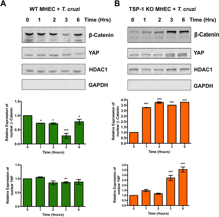
Fig 6. Levels of β-catenin and YAP in nuclear extracts of MHEC during the early phase of T. cruzi challenge.
Nuclear fractions (20 μg) from WT or TSP1 KO MHEC challenged with T. cruzi at different time points were resolved by SDS-PAGE, blotted, and probed with antibodies against β-catenin or YAP in (A) WT MHEC and (B) TSP1 KO MHEC, respectively and developed as described. The blots were stripped, reprobed with antibodies against HDAC1 and developed with the corresponding IRDye conjugated secondary antibody. The blots were further probed with antibodies against GAPDH and developed with the corresponding IRDye conjugated secondary antibody. The developed blots were scanned using the infrared fluorescence detection Odyssey Imaging System. The HDAC1 normalized fold changes in the level of β-catenin and YAP were determined and plotted in the bar graph for WT MHEC (A, middle and lower panel), TSP1 KO MHEC (B, middle and lower panel), respectively. The bar graphs represent mean values ± SE from three independent biological replicates. The value of P< 0.05 was considered significant. *P< 0.05; **P< 0.01; ***P< 0.001.

