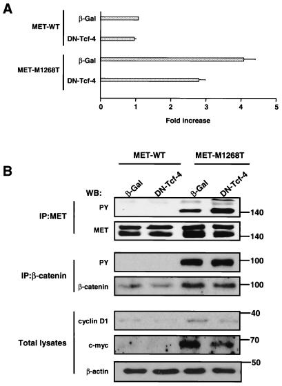
FIG. 7.
(A) MET M1268T receptor mutant causes constitutive transactivation of Tcf consensus sequence (TOPFLASH)-driven transcription in NIH 3T3 cells. Activity of Tcf-4 in NIH 3T3 cells expressing MET WT or M1268T was determined by luciferase assay as described in the legend to Fig. 6A. Luciferase activity in cells expressing MET WT and β-Gal was set equal to 1. Bar graph data are means ± standard errors of three independent experiments. (B) The increased level of c-myc and D1 expression in cells with mutated MET is mediated by Tcf. NIH 3T3 cells expressing MET WT or M1268T were infected with an adenovirus encoding β-Gal or DN Tcf-4. After 48 h the amount of c-myc and cyclin D1 was determined in total lysates from cells by Western blotting with anti-c-myc and anti-cyclin D1 antibodies. MET tyrosine phosphorylation was determined by anti-PY antibodies in MET IPs. To estimate the amount of the MET in precipitates, the blot was probed with anti-MET antibodies (upper band, immature MET [170 kDa]; lower band, mature MET [140 kDa]). Tyrosine phosphorylation of β-catenin was detected by Western blotting (WB) with anti-PY antibodies. The amount of β-catenin in precipitates was determined with anti-β-catenin antibodies. The β-actin panel serves as a control showing an equal amount of protein in each sample. Positions of molecular-weight markers are indicated on the right.

