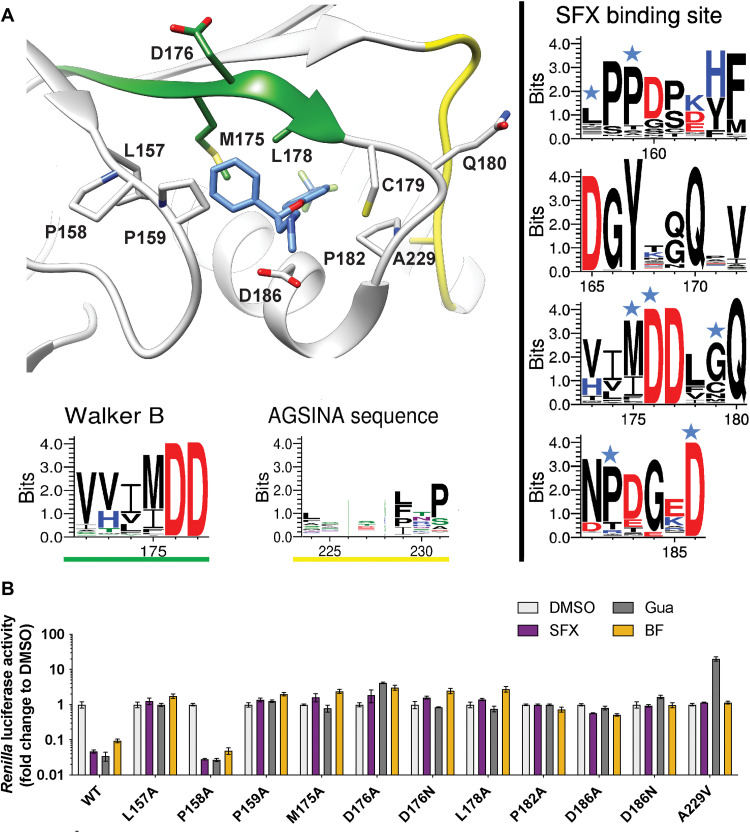
Fig. 2. Mutational analysis of the SFX binding site in CV-B3 2C.
(A) Schematic structure of CV-B3 2C highlighting the residues involved in SFX binding. The Weblogo represents the conservation of amino acids based on an alignment of the 2C protein from EVs that cause disease in humans. The blue stars on top of the amino acid residues in the right panels indicate an interaction with the ligand SFX. (B) The residues that interact with SFX were introduced into an infectious CV-B3 cDNA clone containing the Renilla luciferase reporter gene upstream of the capsid-coding region (Rluc-CV-B3). In vitro transcribed RNA was transfected into cells, and Renilla luciferase was used as a sensitive and quantitative readout for vRNA replication. The experiment was performed three times independently in biological triplicates. One representative experiment is shown.

