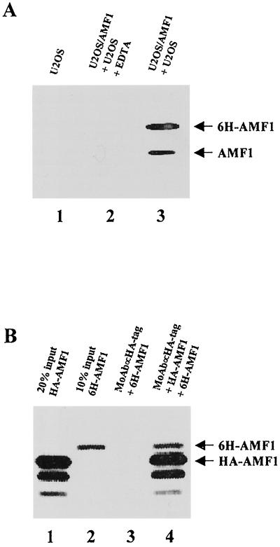
FIG. 6.
Self-association of AMF1 in vivo and in vitro. (A) Coprecipitation of endogenous AMF1 with 6H-AMF1 from U2OS cells. Cell extracts of parental U2OS and U2OS/AMF1 were mixed and incubated with Ni-NTA resin in the presence (lane 2) or absence (lane 3) of 10 mM EDTA. Parental U2OS cell extract alone was also incubated with Ni-NTA resin without EDTA as a control (lane 1). After washing, proteins bound on the resin were eluted and concentrated. AMF1 proteins were detected by Western blotting. (B) In vitro association of HA-AMF1 with 6H-AMF1. Both proteins were prepared by in vitro translation and labeled with [35S]methionine. 6H-AMF1 was incubated with anti-HA MAb 12CA5 (Boehringer Mannheim) and protein A-Sepharose beads with (lane 4) or without (lane 3) HA-AMF1. Protein complexes were resolved on an SDS–15% polyacrylamide gel and analyzed with a Bio-Rad GS-250 molecular imager. Input HA-AMF1 (20%) (lane 1) and input 6H-AMF1 (10%) (lane 2) are shown.

