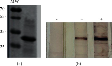
Figure 1.

Polyprotein expression and antigenicity evaluation. Western blot using anti-His (1 : 3,000) as a primary antibody and an alkaline phosphatase-conjugated anti-mouse IgG as a secondary antibody (1 : 3,000). A colorimetric detection was performed using BCIP/NBT (5-bromo-4-chloro-3-indolyl phosphate/nitroblue tetrazolium) Color Development (Promega), according to the manufacturer's instructions (a). The antigenicity of the polyprotein was evaluated in a preparative 12% polyacrylamide gel with the polyprotein solution (100 µl; 40 µg/mL). The strips (∼0.5 cm) from a nitrocellulose membrane with the transferred proteins were exposed for 1 h at room temperature with different bovine sera (1 : 100) of known identity and after the corresponding washes were further incubated for 1 h at room temperature with a phosphatase-conjugated anti-bovine antibody (1 : 5,000), as a secondary antibody. The colorimetric detection was performed using BCIP/NBT as well (b). A representative image is shown. MW: molecular weight; −: negative serum sample; +: positive serum sample.
