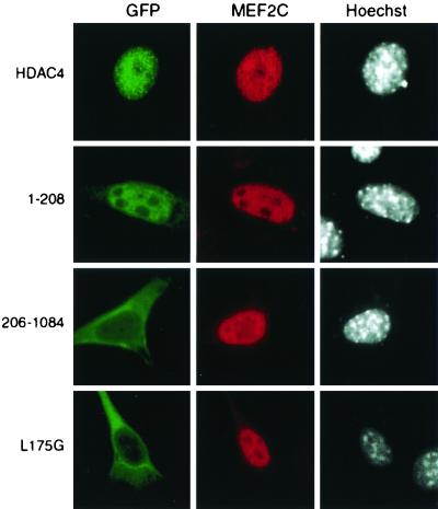
FIG. 9.
Effects of exogenous MEF2C on nuclear localization of HDAC4 and mutants. The MEF2C expression plasmid was transfected into NIH 3T3 cells along with mammalian expression plasmids for GFP fusion proteins of HDAC4 or its mutants. At 16 h after transfection, cells were fixed and stained with anti-MEF2C antibody to detect MEF2C by indirect immunofluorescence microscopy (middle) (red). Green fluorescence was used to determine subcellular distribution of GFP fusion proteins (left) (green). The cells were counterstained with Hoechst 33528 to visualize nuclei (right) (white).

