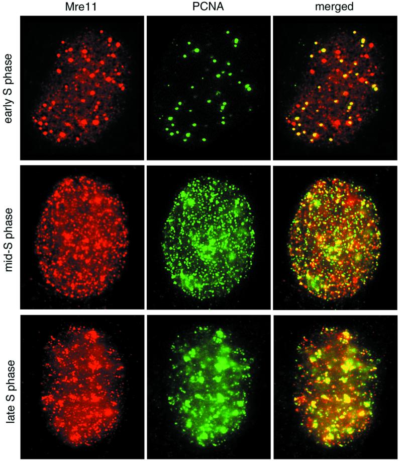
FIG. 6.
Mre11 complex and PCNA colocalization during S-phase progression in normal cells. 37Lu fibroblasts were doubly labeled with Mre11 (red) and PCNA (green) as for Fig. 5b, and the images were merged. Overlap between the two signals in merged images appears yellow. Top row, early S phase; middle row, early- to mid-S phase; bottom row, mid- to late-S phase. Stages were determined by the method of Kill et al. (24).

