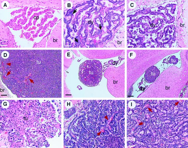
FIG. 2.
Tumor progression in T121p53+/− and T121p53+/+ mice. Normal CP of a 3-month-old nontransgenic mouse (br, brain tissue). Cells contain regularly sized and shaped nuclei with significant cytoplasm in a single layer forming a papillary architecture (A). Due to T121 expression, the CP of young T121p53+/− (3 weeks) (B) and T121p53+/+ (5 weeks) (C) mice appears dysplastic (dy) with nuclei of aberrant sizes and a high nuclear/cytoplasmic ratio in most cells. The overall papillary organization of the tissue is retained; however, multilayered regions are present. The black arrow indicates dysplastic cells, and the pink arrow indicates normal cells. Focal solid masses indicative of tumor (tu) progression are detectable by 6 to 8 weeks in all T121p53+/− mice (7-week-old mouse shown in panel E) and at various times beyond 7 weeks in some T121p53+/+ mice (7-week-old mouse shown in panel F). These focal tumors arise among the background of dysplastic CP. Solid vascularized tumors (designated type I) grow rapidly and become life threatening by 12 weeks in all T121p53+/− mice (12-week-old mouse shown in panels D and H) and at 25 to 43 weeks in 38% of T121p53+/+ mice (38-week-old mouse shown in panel I). The red arrows in these figures indicate blood vessels. Tumors of distinct morphology (type II) also arise in 59% of T121p53+/+ mice (38-week-old mouse shown in panel G). Loosely organized cells within these masses often contain copious cytoplasm and grossly enlarged nuclei. These tumors appear to grow slowly, since they do not reach the large sizes observed for type I tumors. All slides were stained with hematoxylin and eosin. Each white bar represents 50 μm, and each black bar represents 200 μm.

