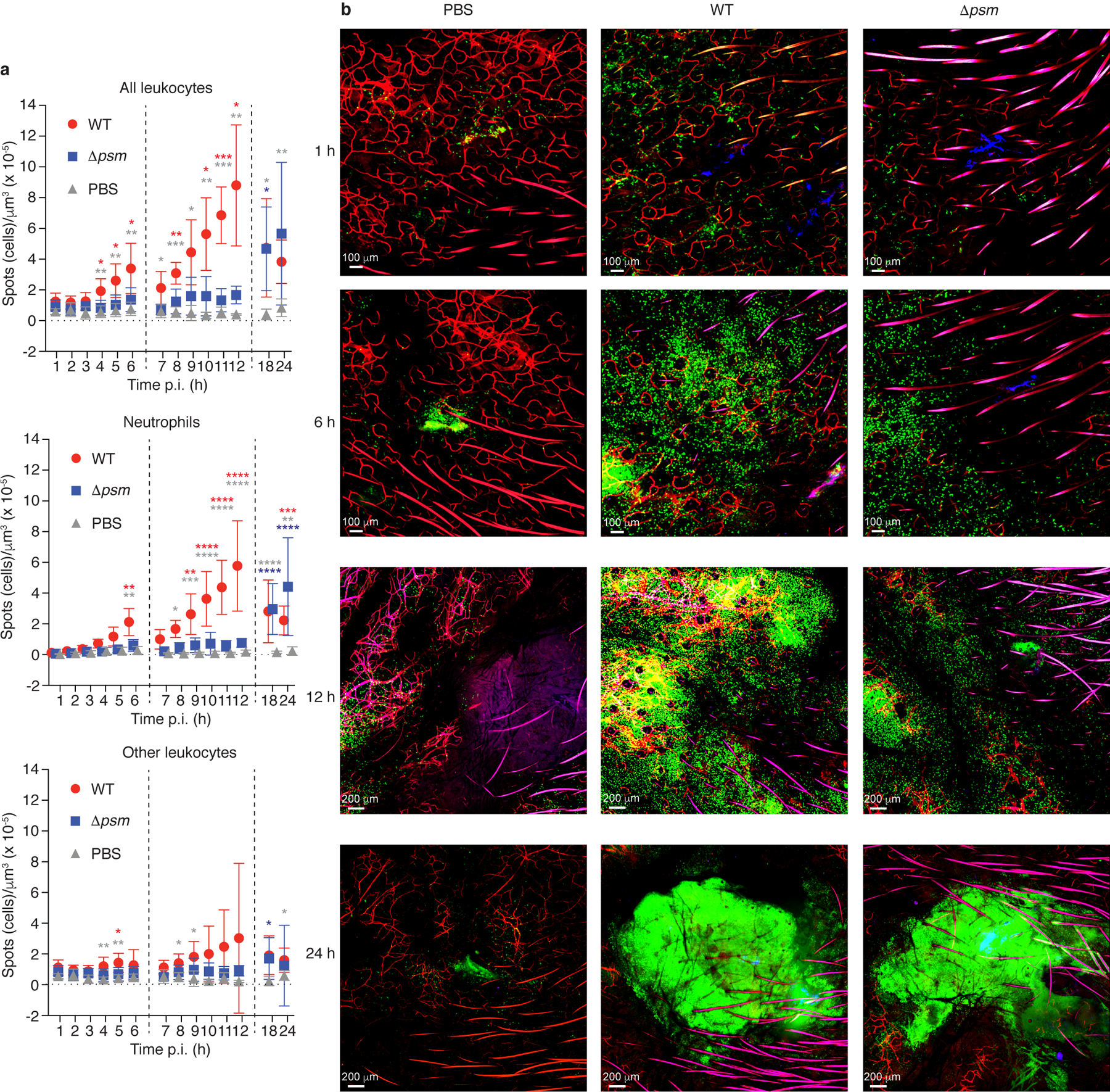
Figure 1. S. aureus PSM toxins are critical to the early leukocyte influx to the site of skin infection - Experiments with bacteria.

a, Influx of total leukocytes, neutrophils, and non-neutrophil leukocytes to the infection site. Data are from the analysis of C-LSM intravital imaging using C57BL/6-Lysozymetm1M-GFP mice. Fluorescently labeled S. aureus LAC (WT) or Δpsm bacteria were injected in a volume of 3 μl into the ear pinnae and images of the infection site were taken every hour over 1 – 6 h and 7 – 12 h, and at 18 h and 24 h p. i. Bacterial inocula were set up to be in the range of 1 – 2 × 10⁷ CFU and actual injected bacterial CFU were determined in the inocula: LAC, 2.48 ± 0.93 (SD) × 107; Δpsm, 2.00 ± 0.85 (SD) × 107 (p=0.21, unpaired two-tailed t-test). Controls received the same volume of PBS. Vertical dashed lines depict separate experiments as mice can only be monitored for a maximum of 6 h. Neutrophils were distinguished from other leukocytes in the image analysis protocol by their higher fluorescence. n = 4 – 8/group. Statistical analysis is by 2-way ANOVA or mixed model with Tukey’s post-tests. *, p<0.05; **, p < 0.01; ***, p < 0.001; ****, p < 0.0001; red asterisks, WT versus Δpsm; grey asterisks, WT versus PBS; blue asterisks, Δpsm versus PBS. Error bars show the mean ± SD. b, Selected representative images. Green, leukocytes; blue, bacteria; red, blood vessels (fluorescent antibody-labeled).
