Fig. 5.

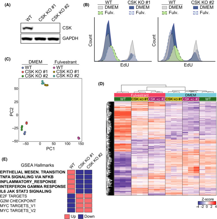
CSK‐dependent growth arrest mediates activation of the inflammatory phenotype in MCF7 cells. (A) Western blot illustrating the loss of CSK expression in 2 independent clones of CSK‐deficient MCF7 generated by CRISPR/Cas9. GAPDH is added as a loading control. (B) Flow cytometric data illustrating the growth arrest that is observed in WT MCF7 cells grown in fulvestrant for 20 days. Two clones of CSK‐deficient cells continue to incorporate EdU in the same conditions. (C) Principal component analysis (PCA) plot of an RNAseq experiment where the transcriptome of two independent MCF7 CSKko clones grown in DMEM with or without fulvestrant (1 μm) for 20 days was compared. The PCA plot was based on normalized gene counts after filtering for low‐expressed genes. (D) Heatmap and clustering of genes from the experiment defined in (C). The genes shown in the heatmap are genes that were found significantly regulated in any of the comparisons. Each gene (row) is standardized (z‐score) to mean = 0 and sd = 1 and then clustered by hierarchical clustering. Note that the fulvestrant‐induced changes in WT MCF7 cells are significantly milder in CSK‐deficient cells. (E) Impact of CSK deficiency on fulvestrant‐dependent expression of genes related to specific GSEA pathways from the experiment defined in (C). The full GSEA is provided in Table S4.
