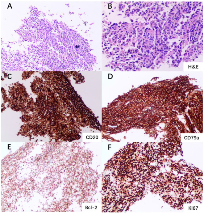
Figure 3.

Left mandible biopsy and immunohistochemistry. (A) Multiple fragments of a squamous mucosa and soft tissue with a dense lymphoid infiltrate (×100 magnification). (B) Aggregate of atypical lymphoid cells (H&E staining; ×200 magnification). (C) Immunohistochemical staining showing sheets of large mononuclear lymphoid cells positive for CD20 (×100 magnification). (D) Sheets of large mononuclear lymphoid cells positive for CD79a (×100 magnification). (E) Cells positive for Bcl-2 staining (×100 magnification). (F) Cells positive for Ki67 staining (×100 magnification).
