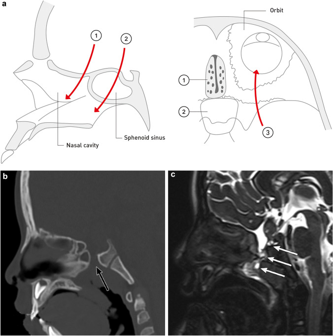
Fig. 6.
Congenital skull base cephalocele. a Classification of congenital skull base cephalocele. Skull base cephaloceles are classified into (i) transethmoidal, (ii) transsphenoidal, and (iii) spheno-orbital. The arrows indicate the pathway by which intracranial contents protrude into the extracranial space through a skull base defect. b Congenital skull base cephalocele (transsphenoidal cephalocele) in a 5-year-old boy with bacterial meningitis and growth hormone deficiency. Sagittal head computed tomography shows a partial defect of the sphenoid bone (black arrow). c Sagittal T2-weighted magnetic resonance image shows herniated contents extended into the nasopharynx through the defect of the sphenoid bone (transsphenoidal cephalocele). Herniated content shows heterogenous intensity, with fluid components showing high intensity, and herniated brain parenchyma and dysplastic gliotic tissue showing iso intensity (white arrows). The pituitary gland has a deformity and is herniating into the sphenoid bone defect

