Fig. 1.
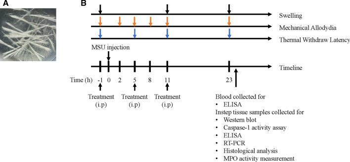
The structure of MSU crystals and experimental protocol. a The structure of MSU crystals in polarizing microscope. b The experimental protocol: MSU crystals (0.5 mg MSU in 20 μL PBS) or PBS (20 μL) was applied into the instep of right paw to establish the acute gouty arthritis model or control group. Swelling was measured at 1 h before MSU injection and 11, 23 h after model establishment. Mechanical allodynia was detected 1 h before MSU injection and 2, 5, 8, 11, 23 h after MSU injection. The measurement of thermal withdraw latency was conducted at 1 h before MSU injection and 5, 11, 23 h after model establishment. Mice were killed to collect blood and instep tissue for Western blot, Caspase-1 activity assay, ELISA, RT-PCR, Histological analysis, and MPO activity measurement

