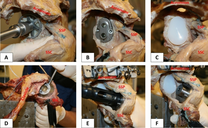
Fig. 3.
Demonstrating the surgical technique. Reaming the articular surface of the glenoid (a). Glenoid guide placed on the central axis of the exposed glenoid surface (b). Inserted keeled glenoid implant (c). Determination of trunnion size on the resected humeral head (d). Insertion of the hollow screw to secure trunnion (e). Trunnion is additionally secured with a small, protruding spike to allow for easy switching of the prosthetic heads (f). SSP = supraspinatus; SSC = subscapularis

