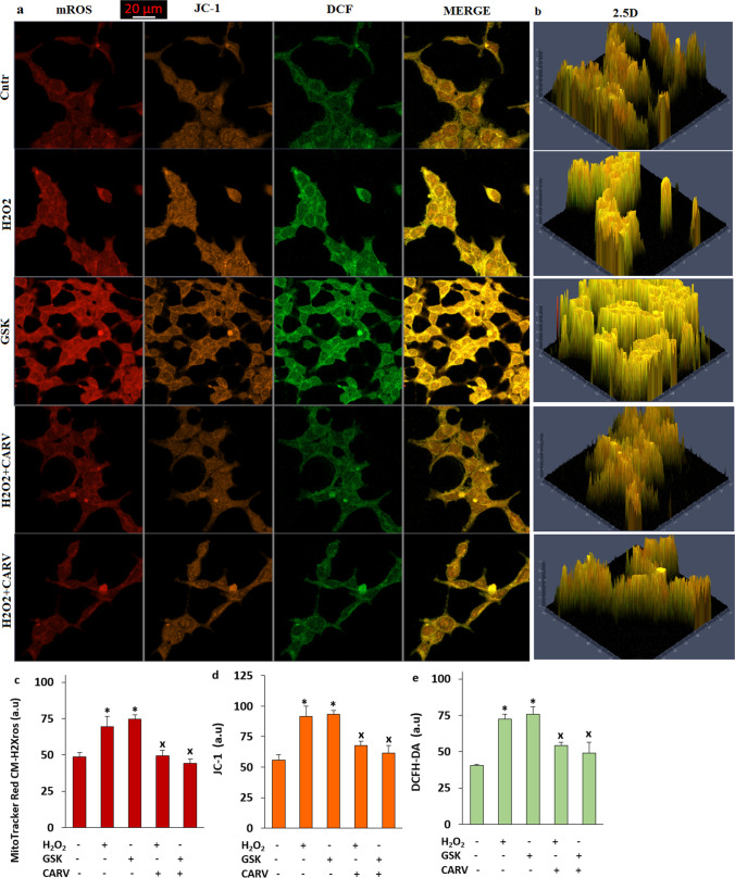
Fig. 9.
The pretreatment of CARV (100 µM for 24 h) diminished GSK (100 nM for 1 h) and H2O2 (1 mM for 1 h)-induced increase of mROS, ΔΨm, and cROS in the SH-SY5Y cells. (Mean ± STD and n = 20-25). a. The images of mROS, ΔΨm, and cROS generation in the Cntr, H2O2, GSK, H2O2+ CARV, and GSK + CARV groups by using the stains of MitoTracker Red CM-H2Xros, JC-1, and DCFH-DA, respectively (Objective: 40×oil. Scala bar: 20 μm). b. The 2.5 images of mROS, ΔΨm, and cROS in the five group. Representative column of the fluorescence intensities of the mROS (c), (d), and DCFH-DA (e). (*p ≤ 0.05 vs. Cntr. ×p≤ 0.05 vs. H2O2 and GSK groups)

