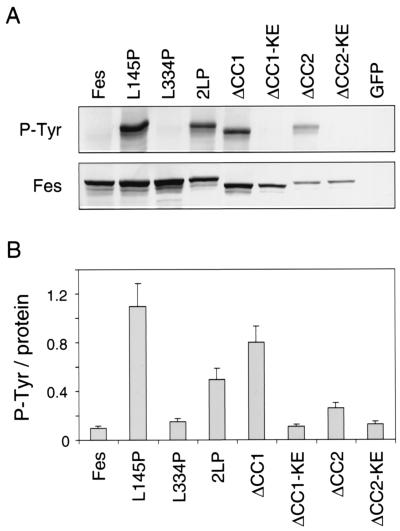
FIG. 3.
Autophosphorylation of GFP-Fes proteins in Rat-2 fibroblasts. Rat-2 fibroblasts stably expressing each of the GFP-Fes constructs shown in Fig. 2 were lysed, and GFP-Fes fusion proteins were precipitated with the M2 anti-FLAG monoclonal antibody. (A) Immunoprecipitates were washed with RIPA buffer, resolved by SDS-PAGE, and immunoblotted for Fes expression with the anti-FLAG antibody (Fes) or for phosphotyrosine content with the antiphosphotyrosine monoclonal antibody PY99 (P-Tyr). A representative blot is shown. (B) The experiment was repeated three times, and the relative levels of GFP-Fes protein and phosphotyrosine content were quantitated using an imaging densitometer. Ratios of tyrosine autophosphorylation to Fes protein were calculated and are presented as means ± standard deviations. KE, kinase-inactive form.

