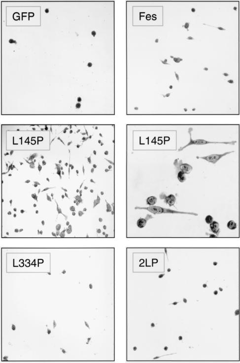
FIG. 7.
A point mutation in the GFP-Fes CC1 domain induces attachment and spreading of TF-1 myeloid leukemia cells. TF-1 cells were infected with a recombinant GFP retrovirus or the indicated GFP-Fes retroviruses and selected as described in the legend to Fig. 6. Following selection, the cells were replated in 24-well plates in the presence of GM-CSF and returned to the incubator for 5 days. Cells were stained in situ with Giemsa stain and visualized by light microscopy. Phase-contrast images of TF-1 cells expressing GFP, GFP-Fes, GFP-Fes L334P, and GFP-2LP were recorded under low-power magnification (×100). Images of cells expressing the GFP-Fes CC1 domain point mutant (L145P) protein were recorded under low-power (middle left panel) and high-power (×400; middle right panel) magnification.

