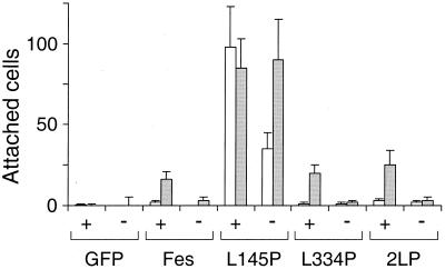
FIG. 8.
Quantitative comparison of the effects of GFP-Fes CC domain mutant proteins on TF-1 cell morphology in the presence and absence of GM-CSF. TF-1 cells were infected with a recombinant GFP retrovirus or the indicated GFP-Fes retroviruses and selected as described in the legend to Fig. 6. For attachment experiments, 5 × 10⁴ cells were plated in duplicate 24-well plates in the presence (+) or absence (−) of GM-CSF. Cells were returned to the incubator for 5 days (open bars) or 7 days (shaded bars), stained with Giemsa stain, and visualized by light microscopy. The number of attached cells was counted in three randomly chosen fields, and results are presented as average values ± standard deviations. This entire experiment was performed twice and produced the same pattern of responses in each case.

