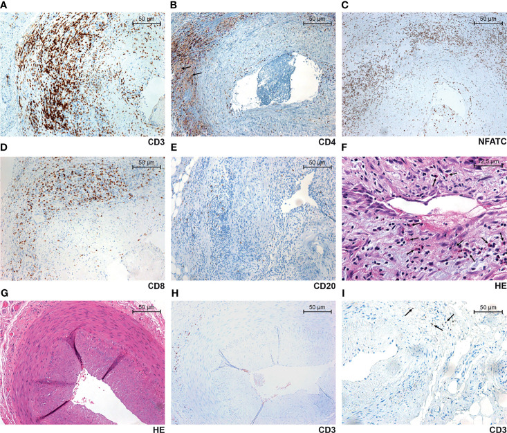
Figure 1.
Selected representative images of TAB-positive GCA arteries (A–F), TAB-negative GCA arteries (G, H) and non-GCA temporal arteries (I). Prominent arterial wall infiltration with CD3+ (A), CD4+ (B) and NFATC+ (C) cells. In addition to strong staining of lymphocytes, there was also a faint CD4 positivity of macrophages and multinucleated giant cells (arrows) (B). In a majority of biopsies, a significant number of CD8+ lymphocytes (D), relatively small number of CD20+ lymphocytes (E), and a variable number of eosinophil granulocytes (arrows) (F) were found. Although not apparently present in hematoxylin and eosin (HE) stain (G), some CD3+ lymphocytes could be found focally segmentally in the adventitia of TAB-negative GCA arteries (H), and a very few in non-GCA temporal arteries of the control group (arrows) (I).

