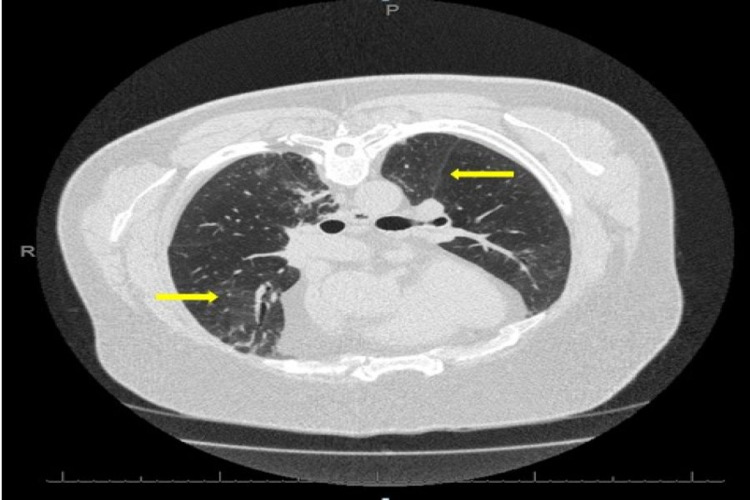
Figure 1. HRCT-axial view.
There is diffuse and patchy ground-glass attenuation (yellow arrows) with tiny nodules in the upper and mid zones, and there are multifocal areas of peripheral consolidation with tractional bronchiolar dilatation within both lower lobes.
HRCT: high-resolution computed tomogram

