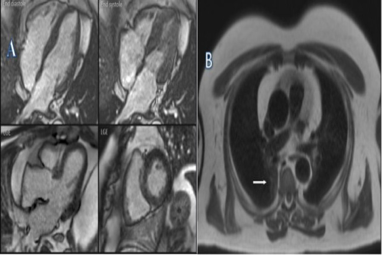
Figure 2. Cardiac MRI.
A. STIR-T2 images: Mild fibrosis of the basal septum and inferior and lateral walls. No myocardial inflammation or infarction. B. Gadolinium study: In the late phase, there is a mild mid-wall enhancement in the basal septum and inferior and lateral walls. There is an area of increased signal in the posterior right lung (white arrow).
STIR: short tau inversion recovery

