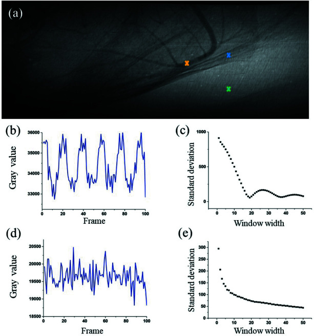
Figure 1.
Standard deviation (SD) of signals introduced by tissue motion and background versus window width. (a) An original projection in the image sequence collected for angiography of the mouse brain. (b) Time-sequenced signal of tissue movements. (c) SD of tissue signal versus window width. (d) Time-sequenced signal of random noise. (e) SD of background noise signal versus window width.

