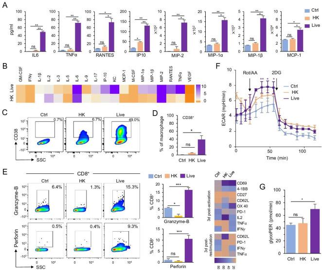
Figure 1.
Live BmΔvjbR treatment activates CD8+ T cells to produce proinflammatory cytokines by polarizing macrophages. (A, B) Cytokine array analysis of the culture medium of BMDMs after treatment with 1× PBS (Ctrl), HK or live) BmΔvjbR for 24 hours shown live BmΔvjbR promotes BMDMs to secret proinflammatory cytokines and chemokines. (C, D) Flowcytometric analysis of CD38 expression on BMDMs after treatment with HK or live BmΔvjbR for 24 hours. (E) Coculturing with live BmΔvjbR-infected BMDMs activates CD8+ T cells to produce granzyme B and perforin (left) and express activation markers and cytokines (right heatmap). (F, G) Cocultivation with live BmΔvjbR-infected BMDMs increases the glycolysis of CD8+ T cells. Data represent means±SD from three independent experiments. *, **, ***Significance at p<0.05, 0.01, and 0.001, respectively. BMDM, bone marrow-derived macrophage; Ctrl, control; ECAR, extracellular acidification rate; GM-CSF, granulocyte-macrophage colony-stimulating factor;HK, heat-killed; IFN-γ, interferon gamma; IL, interleukin; IP-10, interferon gamma-induced protein 10; KC, keratinocytes-derived chemokine; MCP-1, monocyte chemoattractant protein 1; M-CSF, macrophage colony-stimulating factor; MIP, macrophage inflammatory protein; ns, not significant; RANTES, chemokine (C-C motif) ligand 5; VEGF, vascular endothelial growth factor; PBS, phosphate-buffered saline; SSC, side scatter; TNF-α, tumor necrosis factor alpha.

