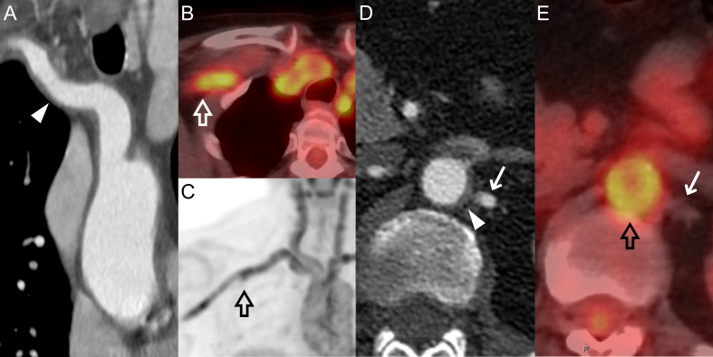
Figure 1.
CTA and PET-CT images of a 64-year-old female patient with LV-GCA. The wall thickening of the innominate and right subclavian arteries (arrowhead) (A) correspond to increased uptake (PET score=3) in the same vascular segments (empty arrows) (B) and (C). The abdominal aorta was also affected by both vessel wall thickening (arrowhead) (D) and increased uptake with PET score=3 (empty arrow) (E), while no thickening or uptake can be seen in the left renal artery (arrows) (D) and (E). CTA, CT angiography; LV-GCA, large vessel-giant cell arteritis; PET, positron emission tomography.

