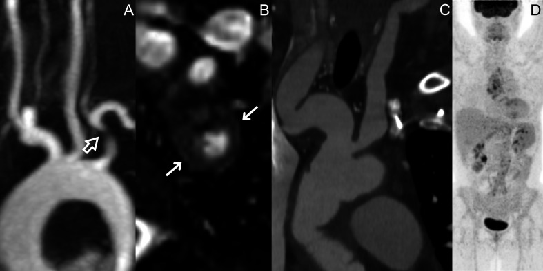
Figure 2.
MRA of a 27-year-old female patient with TAK presenting with a stenosis of the proximal tract of the left subclavian artery (empty arrow) (A), with associated wall thickening in the same segment (arrows) (B). CTA and PET scan of a 53-year-old female patient with LV-GCA who had irregular vascular dilations affecting the innominate artery and the carotid arteries (C), which did not correspond to any 18F-FDG uptake at synchronous PET scan (D). CTA, CT angiography; FDG, fluorodeoxyglucose; LV-GCA, large vessel-giant cell arteritis; MRA, MR angiography; PET, positron emission tomography; TAK, Takayasu arteritis.

