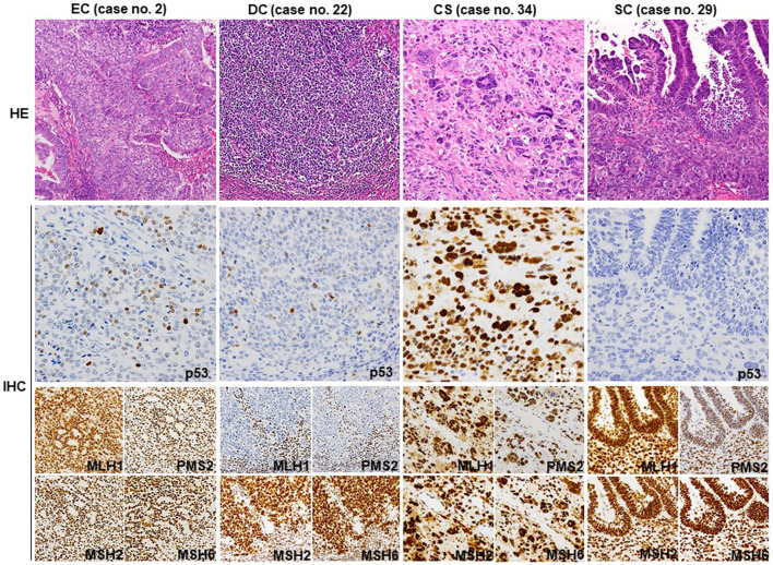
FIGURE 1.
Representative histology and IHC of p53 and MMR proteins. Representative HE sections from G2 EC (no. 2) showing well differentiated glandular and less differentiated solid areas. A few p53-positive tumor cells are observed, indicating wt p53 expression. All four MMR proteins are diffusely positive. The dedifferentiated part of DC (no. 22) exhibits wt p53 expression pattern and loss of MLH1 and PMS2 expression. Stromal lymphocytes also show a positive reaction as an internal control. The sarcomatous element of CS (no. 34) shows diffuse staining for p53 and all four MMR proteins. SC (no. 29) shows complete loss of p53 expression in both glandular and solid elements. The MMR protein expression is well preserved. CS, carcinosarcoma; SC, serous carcinoma; HE, hematoxylin and eosin (Original magnification: ×200); IHC, immunohistochemistry. (Original magnification: p53 × 400, MMR ×200).

