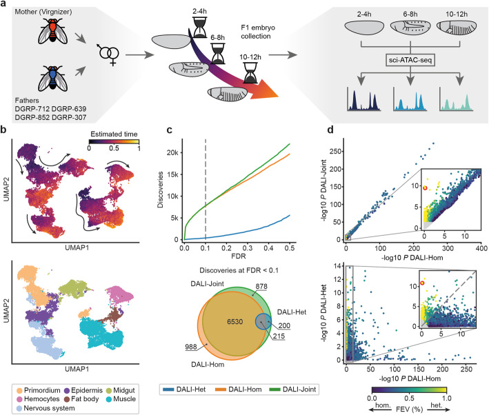
Fig. 2.
Application of scDALI to sciATAC-seq of Drosophila F1 embryos. a Experimental design. Chromatin accessibility was profiled in four F1 crosses at three distinct developmental stages (2–4, 6–8, and 10–12 h after egg laying), resulting in 12 sciATAC-seq libraries. b UMAP visualization of the full integrated dataset (34,053 cells from 12 sciATAC-libraries, excluding cell clusters with ambiguous annotations) based on the latent space of the Variational Autoencoder (VAE) (Methods). Top: Cells colors by the continuous temporal ordering as estimated from the VAE model. Bottom: Colored by the major lineage annotation. c Number of sites across crosses and time points with allelic imbalance identified by scDALI-Joint, scDALI-Hom, and scDALI-Het. Top: Number of discoveries as a function of the FDR threshold (Benjamini Hochberg adjusted). Bottom: Overlap between the sites identified by all three scDALI tests (FDR < 0.1). d Scatter plot of negative log P-values between scDALI-Joint and scDALI-Hom (top) and scDALI-Het versus scDALI-Hom (bottom) respectively. Color denotes the estimated fraction of allele-specific variance explained by cell-state-specific effects; non-significant peaks marked in grey (adjusted scDALI-Joint P > 0.1). Inset plots zoom in on peaks with pronounced cell state-specific imbalances. The red circle highlights the peak chr3R:20310056-20311056, a region showing prominent cell state-specific effects with no discernable global imbalance (c.f. Fig. 3a–c)

