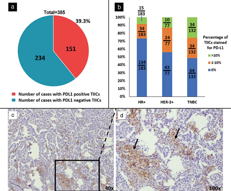
Figure 4.

a) Pie chart representing breast cancer cases (n = 385, after eliminating seven cases in which IHC failed) which had tumor infiltrating immune cells (TIICs) expressing PD-L1 (151/385). b) Column graph depicting the distribution of cases with TIICs stained for PD-L1 (categorized by breast cancer subtypes). c) Lymphoid cells with PD-L1 around tumor cells. d) Magnified image of c showing the infiltration of PD-L1 positive immune cells.
IHC: Immunohistochemistry, PD-L1: Programmed cell death ligand 1
