. 2021 Dec 6;14(2):263–276. doi: 10.1039/d1nr06512b

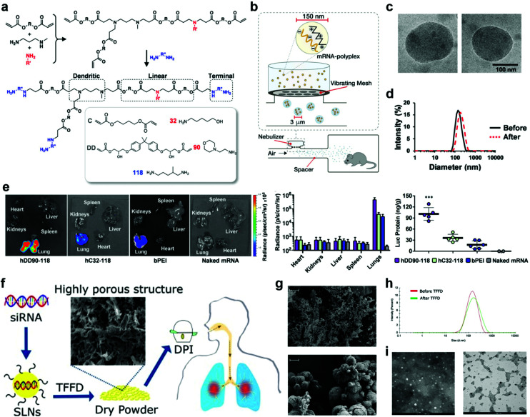
Fig. 3. Pulmonary delivery with different devices and formulations. (a–e) Delivery of liquid formulations with a nebulizer. (a) Synthesis scheme of hDD90-118 and hC32-118 hyperbranched poly(beta amino esters) (PBAEs). (b) A vibrating mesh nebulizer connected to a whole-body chamber was used to deliver in vitro transcribed (IVT)-mRNA encoding for firefly luciferase to mice. The nebulizer generates micrometer sized droplets optimal for lung deposition, containing nanoparticles for intracellular delivery. (c) Electron microscopy of hDD90-118 particles before (left) and after (right) nebulization, particles had an average size of 137 nm (±21) and 146 nm (±40), respectively (±SD, n = 13–15). (d) Particles have narrow size distribution with polydispersity indices of 0.10 before (black) and 0.11 after (red, dashed) nebulization. (e) Bioluminescence 24 h after inhalation of polyplexes, hDD90-118 vectors produced significantly higher level of radiance (±SD, n = 4) and luciferase production (***P < 0.001, ±SD, n = 5–6) in the lungs, compared to hC32-118 and bPEI. Statistical analysis using one-way ANOVA with post-hoc Tukey test. Reproduced with permission.19 Copyright 2019 WILEY-VCH Verlag GmbH & Co. KGaA, Weinheim. (f–i) Delivery of dry powder formulations with a dry powder inhaler (DPI). (f) Schematic illustration of the preparation, thin-film freeze-drying (TFFD), and pulmonary delivery of aerosolizable siRNA-encapsulated solid lipid nanoparticles (LNPs). (g) Representative scanning electron microscope (SEM) images of dry powders of LNPs prepared by TFFD (upper panel) or spray drying (lower panel). The scale bar indicates 2 μm. (h) A comparison of siRNA-encapsulated LNPs particle size distribution by intensity. (i) Transmission electron microscopy (TEM) images of siRNA-encapsulated LNPs before (left panel) and after (right panel) they were subjected to TFFD and reconstitution. Reproduced with permission.32 Copyright 2021, Elsevier B.V.

Fig. 3