
Figure 3. Somatosensory and anterior cingulate intraneocortical connections (INC) in postnatal day 1 (P1) pups.
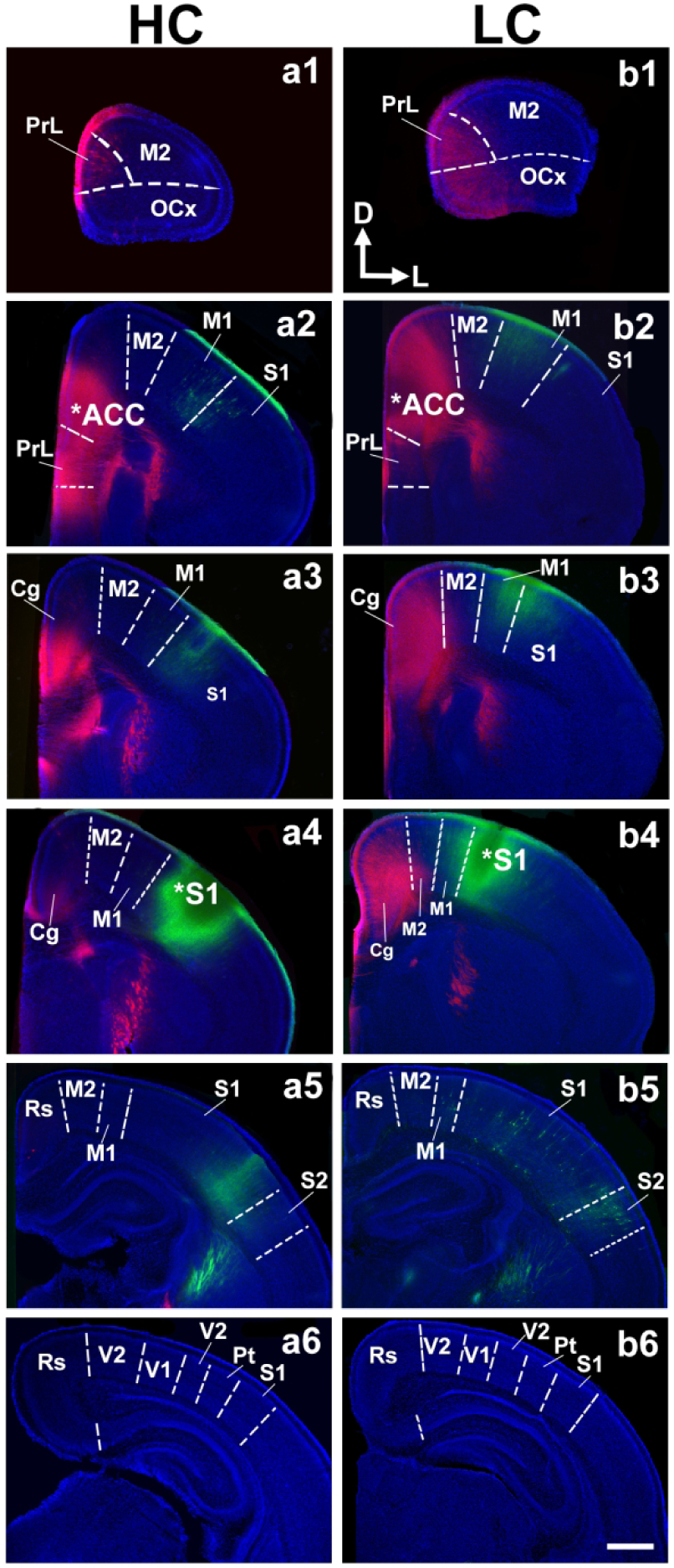
Vibratome-cut 100 μM coronal sections from P1 hemispheres are arranged in a rostral (top) to caudal (bottom) series following crystal placement of DiI (red) or DiA (green) in putative anterior cingulate cortex (ACC; a2, b2, stars) and somatosensory (S1; a4, b4, stars) cortex of high contact (HC; a1–6), and low-contact (LC; b1–6) offspring brains. Sections were counterstained with DAPI (blue). Analysis of ACC and S1 connections revealed no difference between LC and HC offspring. Sections are oriented dorsal (D) up and lateral (L) to the right. Scale bar = 500 μm.