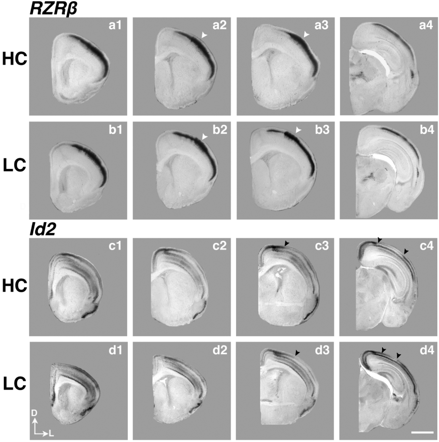
Figure 6. Analysis of neocortical expression of RZRβ and Id2.

High magnification P1 coronal sections of high-contact (a1–4) and low-contact (b1–4) offspring in situ hybridized to RZRβ, as well as sections of high-contact (c1–4), low-contact (d1–4) offspring in situ hybridized to Inhibitor of DNA binding 2 (Id2). White arrowheads (a2–3, b2–3) indicate strong RZRβ expression in the developing barrel cortex in S1, similar to expression patterns in mouse development. Black arrowheads in c3–4 and d3–4 indicate the border of superficial Id2 expression, which is shifted laterally in LC pups compared to HC pups. Images oriented dorsal (D) up, lateral (L) right. Scale bar = 1,000 μm.
