
Figure 8. Analysis of RZRβ and Id2 expression at the sensory-motor border.
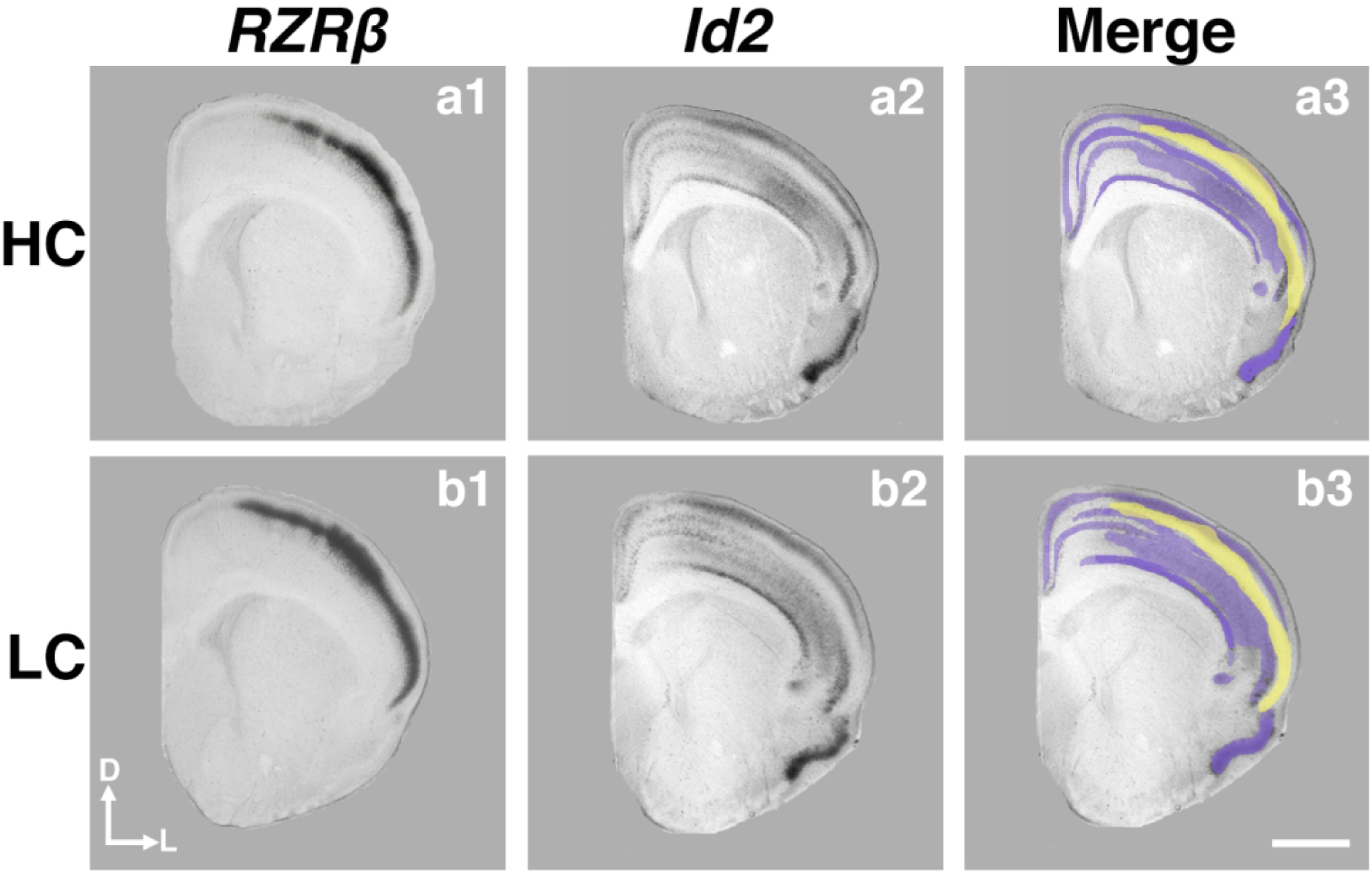
P1 coronal sections of HC (a1–3) and LC (b1–3) offspring hybridized to RZRβ (a1, b1) and Id2 (a2, b2) at the level of the somatosensory-motor cortex. Merging the expression patterns together (a3, b3) reveals the primarily complementary patterning of RZRβ and Id2 expression at this level, as well as a thin overlapping region in both LC and HC offspring. Purple, Id2 expression; yellow, RZRβ expression. Images oriented dorsal (D) up, lateral (L) right. Scale bar = 1,000 μm.