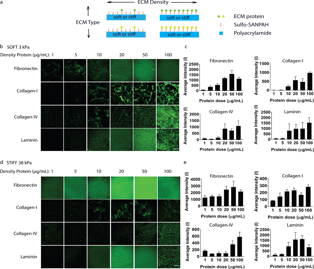
Figure 1: Characterizing ECM protein incorporation with varying type on hydrogel substrates.
(a) Schematic of experimental set up, (b) incorporation of various ECM proteins with tunable density on 3 kPa soft and (d) 38 kPa stiff hydrogels visualized with immunohistochemistry for the corresponding protein (first row, green: fibronectin; second row, green: collagen-I; third row, green: collagen-IV; fourth row, green: laminin) (Scale bars: 30 μm) and (c) quantification on 3 kPa soft and (e) 38 kPa stiff hydrogels of fluorescence intensity of immunohistochemistry staining of hydrogels coated with various doses of fibronectin (top left), collagen-I (top right), collagen-iv (bottom left), or laminin (bottom right).

