
Figure 1.
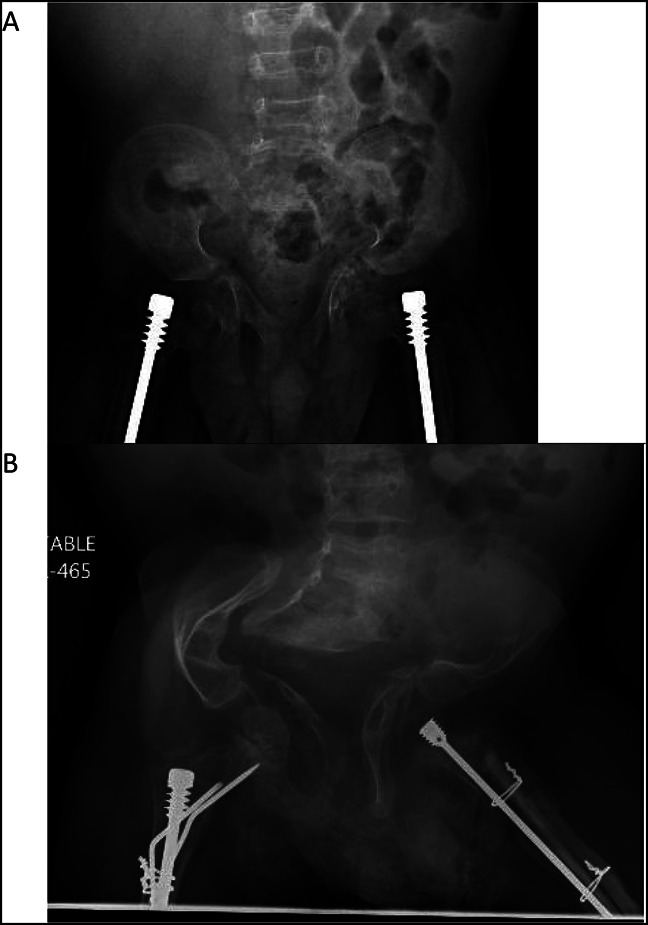
Radiograph of A, AP pelvis at 2 years and (B) AP pelvis at 8 years demonstrating marked progression of the patient's pelvic deformation and acetabular protrusio.
Official websites use .gov
A
.gov website belongs to an official
government organization in the United States.
Secure .gov websites use HTTPS
A lock (
) or https:// means you've safely
connected to the .gov website. Share sensitive
information only on official, secure websites.
Radiograph of A, AP pelvis at 2 years and (B) AP pelvis at 8 years demonstrating marked progression of the patient's pelvic deformation and acetabular protrusio.