Figure 2.

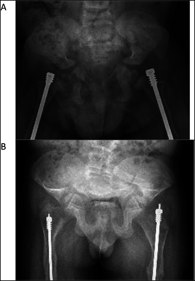
Radiograph of A, AP pelvis at 2 years and (B) AP pelvis at 10 years demonstrating marked progression of the patient's pelvic deformation and acetabular protrusio.

Radiograph of A, AP pelvis at 2 years and (B) AP pelvis at 10 years demonstrating marked progression of the patient's pelvic deformation and acetabular protrusio.