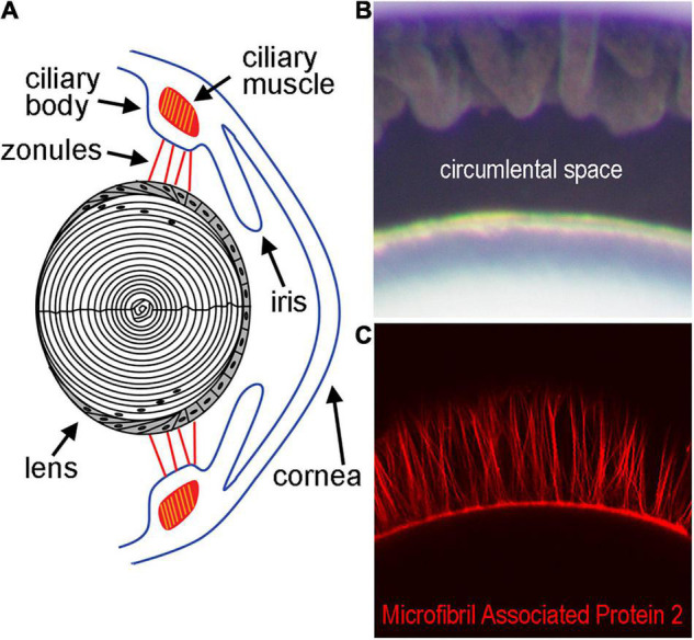
FIGURE 1.

Diagram of the lens and supporting structures. (A) The anterior surface of the lens rests directly behind the iris and is attached by the Zonules of Zinn to the ciliary body. The Zonules of Zinn transduce tension generated by the ciliary muscle to the lens equator during the process of accommodation. (B) The area between the lens and ciliary body containing the zonules in a mouse eye, also known as the circumlental space, as visualized by light microscopy. (C) Fluorescent labeling of the zonules using an antibody against Microfibril Associated Protein-2 visualized by confocal microscopy.
