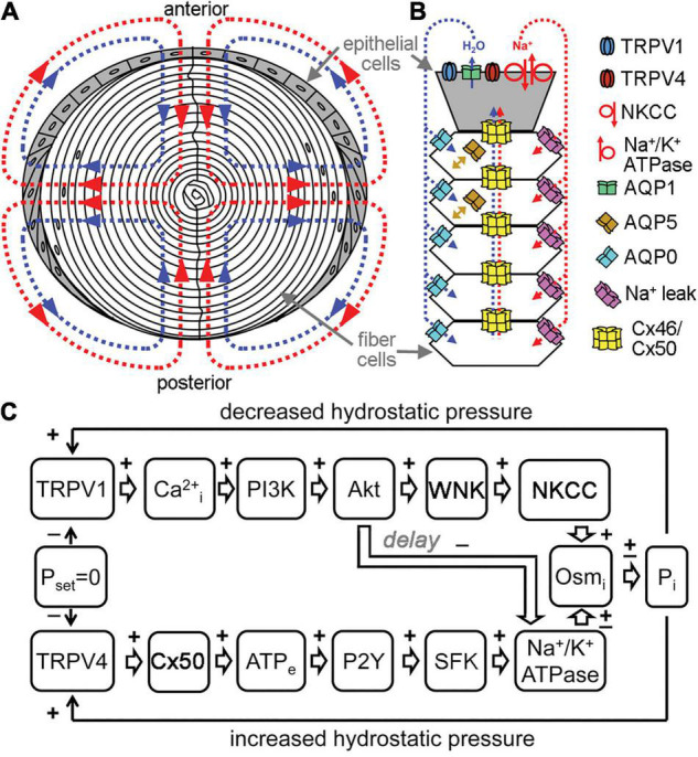
FIGURE 2.

Channels regulate lens transport. (A) The flux of Na+ (red), followed by water (blue), enters the lens at both poles and exits at the equator and acts as a microcirculatory system. (B) Na+ flows into the lens though extracellular spaces, moves into fiber cells through Na+ leak channels, and flows back to the surface through gap junctions, where the Na+/K+-ATPase pumps it out of the lens. Water enters the lens through the extracellular spaces, moves into fiber cells through AQP0 and AQP5 driven by local osmotic gradients created by the transmembrane Na+ flux, and leaves the lens through AQP1 resulting from local osmotic gradients generated by the Na+/K+-ATPase. Hydrostatic pressure drives the water from cell to cell through gap junctions. (C) A feedback control mechanism maintains hydrostatic pressure (P) and water transport in the lens. Decreased pressure activates TRPV1, which then up regulates the NKCC and down regulates the Na+/K+-ATPase through a PI3K/Akt dependent pathway. Increases in pressure activate TRPV4, which then increases Na+/K+-ATPase activity through Cx50, ATP release and purinergic receptor activation of a Src family kinase (SFK).
