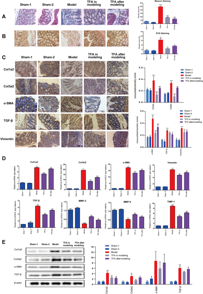
FIGURE 2.
TFA inhibits fibrotic changes in the colon of TNBS-treated mice; (A) Masson’s trichrome staining; (B) Verhoeff’s von Gieson (EVG) staining assay. (×100). (C) Immunohistochemical staining for fibrosis-related proteins in mice from different groups. TNBS induction could significantly increase the expressions of fibrosis-related collagens (col1a2, col3a2), TGF-β, smooth muscle actin (α-SMA), and vimentin, and TFA treatment in modeling or after modeling could reverse the phenomenon. (D) The mRNA expression of fibrosis-related proteins was determined using quantitative real-time PCR. (E) The fibrosis-related protein expression was detected by Western blot analysis. Data are shown as the mean ± SEM. *p < 0.05, **p < 0.01, and ***p < 0.001compared with sham-1 and sham-2 groups, # p < 0.05, ## p < 0.01, ### p < 0.001 compared with the model group.

