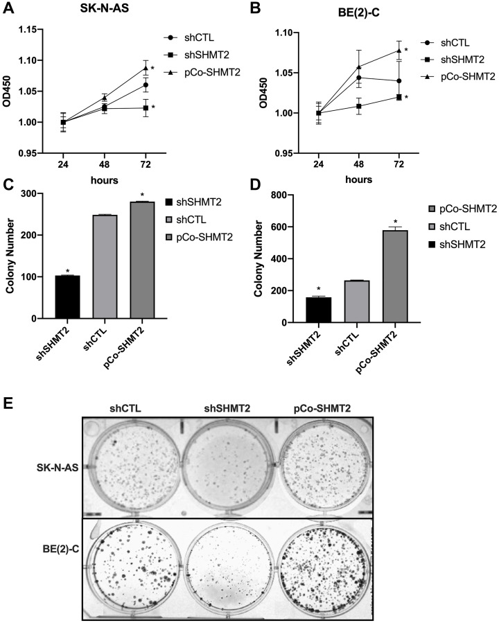
Figure 4. SHMT2 increases cellular proliferation and colony formation in vitro.
(A) SK-N-AS cells were plated on a 96-well plate (1000 cells/well) and cellular proliferation was assessed using CCK-8 assays at 24, 48 and 72 hours after plating. SHMT2 silencing (shSHMT2) decreased cellular proliferation by 1-fold at 72 hours in SK-N-AS cells and SHMT2 overexpression (pCo-SHMT2) increased cellular proliferation by 1-fold at 72 hours. (B) BE(2)-C cells plated onto a 96-well plate (500 cells/well) demonstrated increased cellular proliferation by 1-fold at 72 hours with SHMT2 overexpression and decreased cellular proliferation by 1-fold at 72 hours with SHMT2 silencing, compared to control. (C) SK-N-AS cells were plated on a 6-well plate at 1000 cells/well for 14 days. Cells were stained with 0.05% crystal violet and colony number was counted. SHMT2 silencing (shSHMT2) resulted in a 2.4-fold decrease in mean colony number compared to control (shCTL). SHMT2 overexpression (pCo-SHMT2) cells demonstrated a 1-fold increase in mean colony number compared to control. (D) BE(2)-C cells plated on a 6-well plate at 500 cells/well for 14 days demonstrated a 2.2-fold increase in colony formation with SHMT2 overexpression and 1.7-fold decrease in colony formation with SHMT2 silencing. (E) Colony staining with 0.05% crystal violet demonstrates decreased colony formation in SHMT2 silenced cells (shSHMT2) and increased colony formation with SHMT2 overexpression (pCo-SHMT2) compared to control (shCTL). * p < 0.05.

