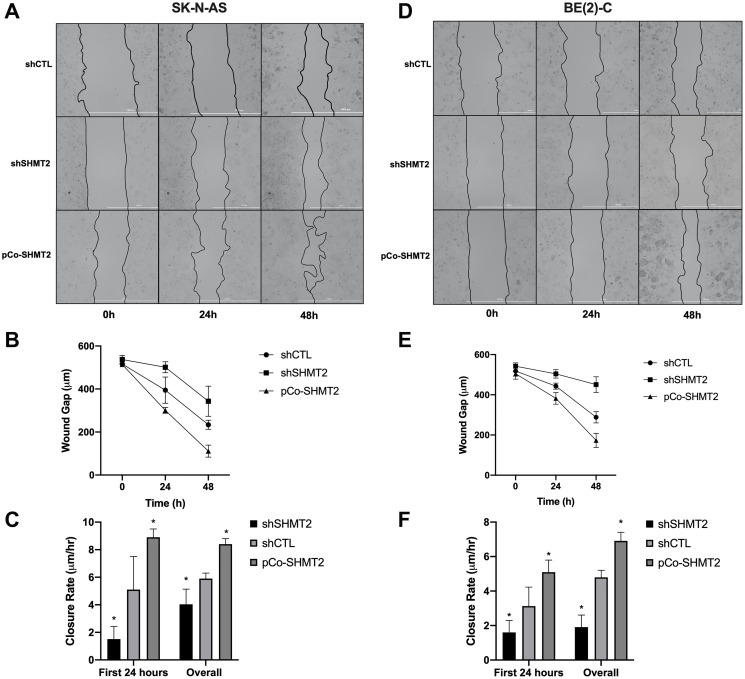
Figure 5. SHMT2 silencing impairs cellular migration in NB.
Cells were plated at varying densities (SK-N-AS at 6.0 × 10⁵ cells/mL and BE(2)-C at 2.5 × 10⁵ cells/mL) in 2-well culture inserts (Ibidi). Once 100% confluency was reached, the cell insert was removed, creating an approximately 500 μM gap. Images were obtained using light microscopy at 0, 24 and 48 hours after wound creation. (A) SK-N-AS cells at 0, 24 and 48 hours. (B) SHMT2 silencing (shSHMT2) increased the average wound gap of SK-N-AS cells and SHMT2 overexpression (pCo-SHMT2) decreased the average wound gap at 0, 24 and 48 hours. (C) SHMT2 silencing decreased the first 24-hour mean wound closure rate by 3.4-fold and overall mean wound closure rate by 1.5-fold, while SHMT2 overexpression increased both the first 24 hour and overall mean wound closure rates by 1.7-fold and 1.4-fold, respectively. (D) BE(2)-C cells at 0, 24 and 48 hours. (E) SHMT2 silencing (shSHMT2) increased the average wound gap of BE(2)-C cells and SHMT2 overexpression (pCo-SHMT2) decreased the average wound gap at 0, 24 and 48 hours. (F) SHMT2 silencing decreased the average wound closure rate at day 1 by 2.0-fold and overall by 2.5-fold, while SHMT2 overexpression increased both the day 1 and overall wound closure rates by 1.6-fold and 1.4-fold, respectively. * p < 0.05.

