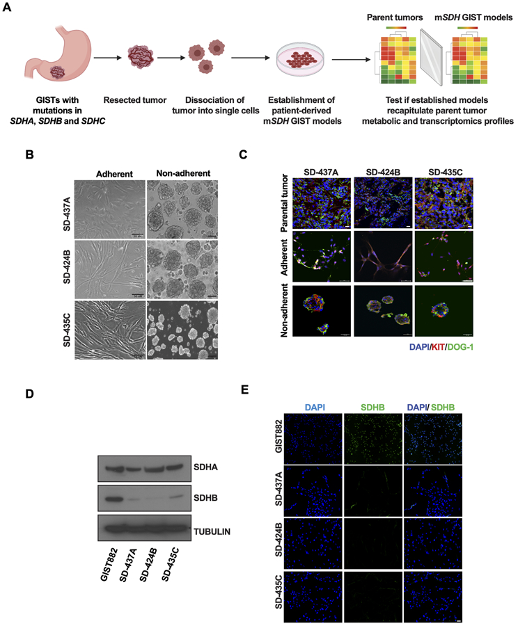
Figure 1. Establishment and characterization of SDHA, SDHB, and SDHC-mutant human GIST models.
A. Schematic representation of workflow of establishment of patient-derived mSDH GIST models and validation for recapitulation of essential features of parent tumors.
B. Micrographs of mSDH GIST models SD-437A, SD-424B, and SD-435C propagated in adherent conditions on a laminin-rich HTB9 matrix (2D) or in non-adherent conditions for 7 days as spheroids on Poly-HEMA coated wells (3D). Scale bar, 50 μm.
C. Immunofluorescence staining of KIT (red) and DOG-1 (green) in parent tumors and mSDH GIST models grown in adherent and in non-adherent conditions.
D. Immunoblot analysis confirming expression of SDHA and SDHB protein in KIT-mutant/SDH-WT (wildtype) GIST882 cells and in mSDH GIST models. α-tubulin was used as a loading control.
E. Immunofluorescence staining of SDHB (green) and DAPI (blue) in KIT-mutant/SDH-WT (wildtype) GIST882 cells and in mSDH GIST models. Scale bar, 50 μm.

