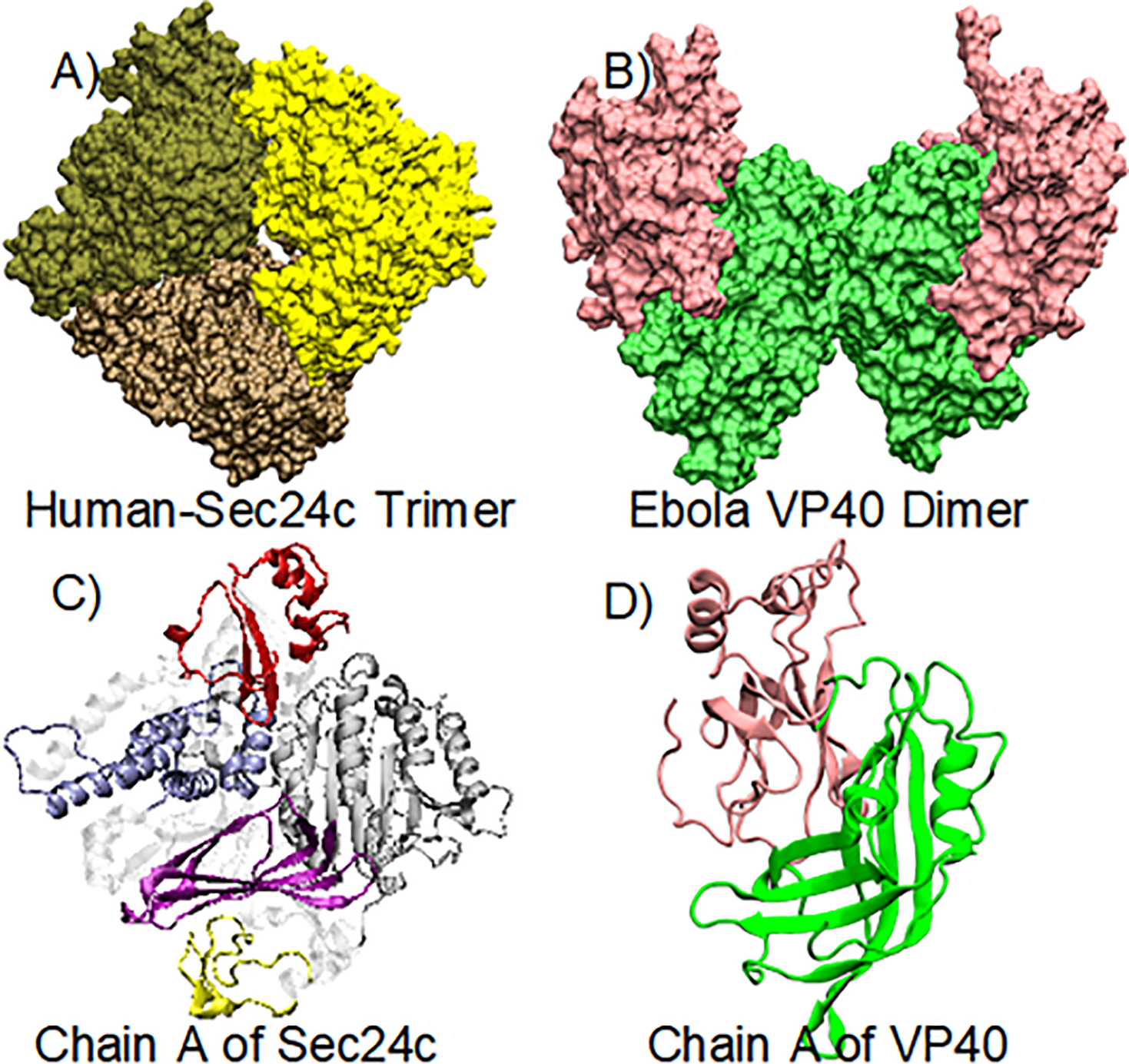
Figure 1.

(A) Crystal structure of the human Sec24c trimer (PDB: 3EH2); the three chains are colored differently. (B) Crystal structure of the Ebola virus VP40 dimer: NTDs (green) and CTDs (pink). (C) Monomer of Sec24c: zinc finger (yellow), trunk domain (gray), beta-sandwich (purple), helical-domain (blue), gelsolin-like domain (red). (D) Monomeric structure of the VP40 protein.
