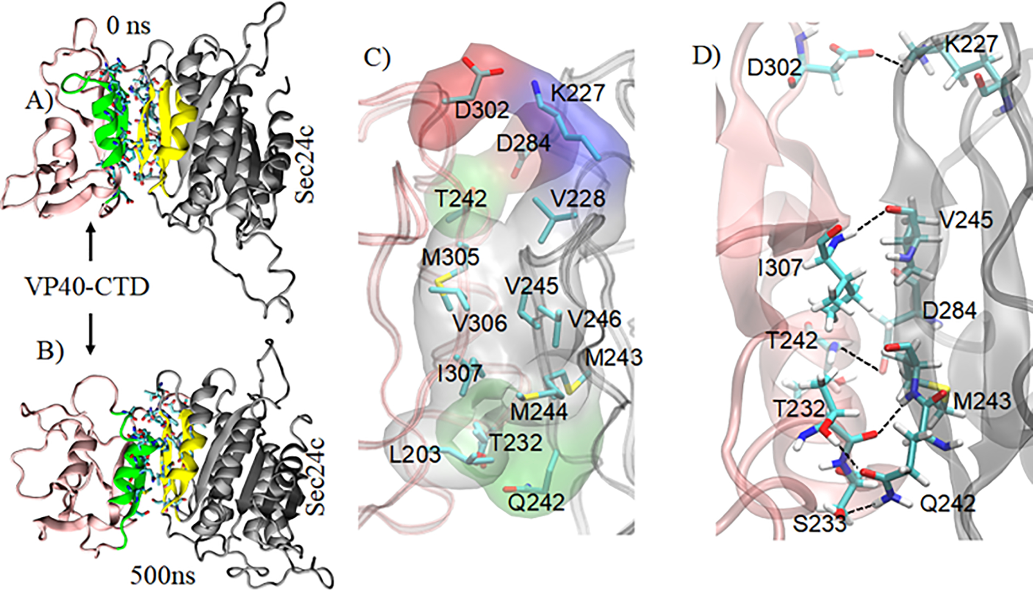
Figure 8.

Sec24c-VP40-AD complex with highlighting of the VP40 binding region (green) and the Sec24c binding region (yellow). MD structure of the complex at: A) 0 ns, B) 500 ns. C) Important residues at the protein-protein interface. Hydrophobic residues (gray) form a strong hydrophobic core that contributes to the stability of the complex. The polar, negative, and positive residues at the interface are shown in green, red, and blue surface representation, respectively. D) View showing some interprotein residue-residue interactions between VP40 and Sec24c.
