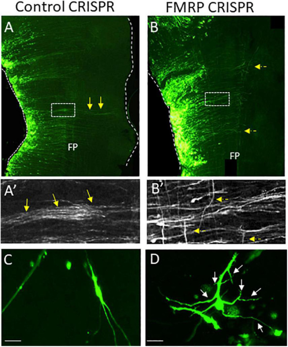
FIGURE 8.

CRISPR-mediated FMRP knockout induces disoriented axonal growth in dA1-derived NM neurons in the chick hindbrain. (A–B′) Flat-mounted hindbrains from embryos electroporated with control (A) or FMRP (B) CRISPR/Cas9- guide RNA-GFP plasmids. Electroporated NM axons are GFP-labeled. Higher-magnification views of the boxed areas in (A,B) are shown in (A′,B′). Yellow arrows indicate aligned axons that cross the hindbrain midline (A,A′). Dashed yellow arrows indicate disoriented axons (B,B′). (C,D) Cell cultures from GFP-expressing NM neurons that were electroporated with the above-mentioned plasmids. Control cells (C) project straight and oriented axons. FMRP-knockout cells (D) project over-branching axons Scale bars: 50 μm. FP, floor plate; NM, nucleus magnocellularis; CRISP, Crisper/Cas9-based plasmids.
