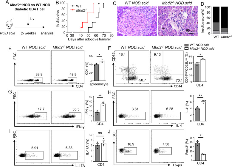
Fig. 3. Adoptive transfer of Mbd2 deficient diabetic CD4 T cells accelerates type 1 diabetes in NOD.scid mice.
A CD4 T cells isolated from newly onset diabetic WT or Mbd2−/− NOD mice were adoptively transferred into 5-week-old NOD.scid recipients (2 × 10⁶ cells/mouse) and the mice were monitored for B diabetes occurrence (n = 6 per group). Pancreases were fixed and processed for C H&E staining and the assessment of D insulitis severity (6 mice per group). Splenic cells were harvested 52 days post adoptive transfer and stained for flow cytometry. Frequencies of E CD4+, F CD4+ CD44high, G CD4+ IFN-γ+ (Th1), H CD4+ IL-4+ (Th2), I CD4+ IL-17A+ (Th17), and J CD4+ Foxp3+ (Treg) cell subsets are shown as representative dot plot graphs. Diabetes incidence B was analyzed by Log-rank test (data were pooled from two independent experiments); insulitis score D was determined by χ2 test, and the flow cytometry results were analyzed by the unpaired Student’s t test (three mice per group with two independent experiments). Data are presented as mean ± SEM. *p < 0.05, **p < 0.01. ns not significant.

