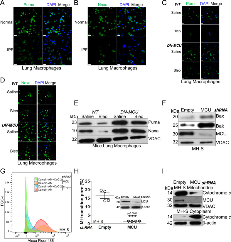
Fig. 2. MCU is associated with apoptosis resistance and inhibition of the mitochondrial intrinsic pathway.
Lung macrophages from normal or IPF subjects were stained and imaged for A Puma or B Noxa by confocal microscopy. Scale bars, 20 μm. Lung macrophages from bleomycin- or saline-exposed DN-MCU-Lyz2-cre mice or WT littermates were stained and imaged for C Puma or D Noxa by confocal microscopy. Scale bars, 20 μm. E Lung macrophages from DN-MCU-Lyz2-cre mice or WT littermates were subjected to mitochondrial isolation and immunoblot analysis for Puma and Noxa. MH-S cells were transfected with empty or MCU shRNA. F Immunoblot analysis for Bax and Bak in isolated mitochondria, and G mitochondrial permeability transition pore opening were determined in live cells by flow cytometry, and H quantified, n = 4. I Macrophages were transfected with empty or MCU shRNA. Immunoblot analysis for cytochrome c was performed in isolated mitochondria and cytoplasm. Two-tailed student’s t-test. **p ≤ 0.01. See also Fig. S2.

