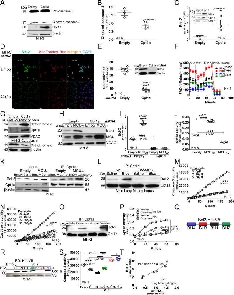
Fig. 3. MCU modulated binding of Cpt1a with Bcl-2 to induce apoptosis resistance.
MH-S cells were transfected with empty or Cpt1a. Immunoblot analysis for A caspase-3 with B statistical quantification, n = 3, (C) Bcl-2 in isolated mitochondria with statistical quantification, n = 3. D MH-S was transfected with Cpt1a shRNA plasmid or empty vector. Cells were stained with MitoTracker Red and Bcl-2 24 h later and subjected to confocal imaging. E The colocalization of Bcl-2 to MitoTracker Red in D was quantitated, n = 3. F MH-S was transfected with Cpt1a shRNA plasmid or empty vector, and cultured for 24 h. Cells were subjected to FAO measurement by Seahorse assay, n = 4–6. G MH-S was transfected with Cpt1a plasmid or vehicle. Cytochrome c in isolated mitochondria and cytoplasm was detected by immunoblot analysis. Macrophages were co-transfected with empty or MCUWT with empty or Cpt1a shRNA. Immunoblot analysis for H Bcl-2 with I statistical quantification, n = 3. J MH-S cells were transfected to overexpress MCUWT, MCUDN, or empty vector. Cpt1a activity was measured, n = 4. K MH-S cells were co-transfected empty or MCUWT in combination with empty or Cpt1a. Cpt1a was immunoprecipitated and immunoblot analysis for Bcl-2 and Cpt1a was performed. L DN-MCU-Lyz2-cre mice and WT littermates were exposed to saline or bleomycin. Lung macrophages were isolated at 21 days, subjected to Cpt1a immunoprecipitation, and immunoblot analysis for Bcl-2 and Cpt1a. M MH-S was treated with octanoate at various concentrations for 3 h. Whole lysate was prepared for determining caspase-3 activities, n = 4. N MH-S was treated with palmitate at various concentrations for 3 h. Caspase-3 activities were measured, n = 4. O MH-S was treated with octanoate (10 µM) or palmitate (100 µM) for 3 h. Whole lysate was precipitated with Cpt1a antibody, and elutes were subjected to detection of Bcl-2 and Cpt1a by immunoblot analysis. P MH-S was treated with octanoate (10 µM, 4 h), in combination with malonyl CoA (100 µM, 3 h) or vehicle. Cell lysate was prepared for quantitation of Cpt1a activities, n = 4. Q Schematic of V5-His tagged Bcl-2 with four BH domains. R MH-S cells were transfected with Bcl-2-V5-His full length or truncations of BH1, BH2, BH3, or BH4. Bcl-2-V5-His was purified by pull down and immunoblot analysis for Cpt1a was performed. S MH-S cells were transfected with empty or Bcl-2-V5-His constructs. Caspase-3 activity was performed, n = 4. T Pearson’s correlation of CPT1A and Bcl-2 expression in IPF lung macrophages. One-way ANOVA with Tukey’s post hoc comparison (I, J, M, N, S). Two-tailed student’s t-test (B, C, E). *p ≤ 0.05, **p ≤ 0.01, and ***p ≤ 0.001. See also Fig. S3.

