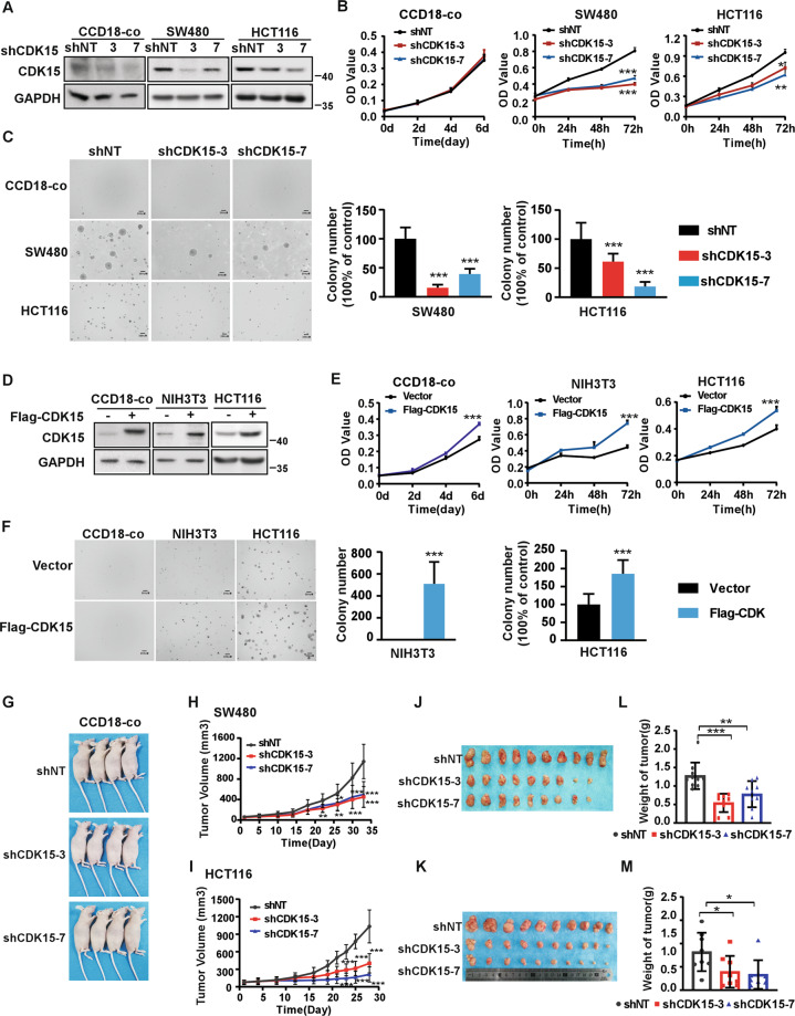
Fig. 2. CDK15 promotes colorectal cancer cell growth in vitro and in vivo.
A, D Cells with CDK15 knockdown and overexpression were established and the expression of CDK15 was determined by western blot. B, E Cell proliferation was measured by MTT assay. C, F Anchorage-independent growth from different cells with CDK15 knockdown or overexpression. Colonies were counted using Image J-Plus (Scale bar: 200 μm). Data represent statistical analysis of colony number in right panel. G CCD18-co cells stably infected with lentivirus (shNT, shCDK15-3, shCDK15-7) subcutaneously were injected into the right flank of nude mice (n = 4 mice per group). H, I SW480 and HCT116 cells stably infected with lentivirus (shNT, shCDK15-3, shCDK15-7) were subcutaneously injected into the right flank of nude mice (n = 9–10 mice per group). Tumor volume was monitored every 2–5 days. J, K Tumor photographs. L, M Tumor weight. Data from in vitro experiments were presented as means ± SD from triplicate experiments. All data statistical differences were evaluated using Student’s t-test. *P < 0.05, **P < 0.01, ***P < 0.001.

