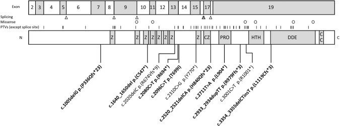
Fig. 1. Exon and protein models of POGZ showing location of the variants.
On top is an exonic model of POGZ (the first exon is non-coding). Below is the POGZ protein which contains a series of zinc finger domains (Z), a chromobox 5 binding region which contains a further zinc finger domain (CZ), proline-rich domain (PRO), helix-turn-helix centromere protein-B-like DNA-binding domain (HTH), a DDE superfamily endonuclease domain (DDE) and a coiled coil domain (CC). Below the protein model are variants reported in this series. Novel variants are bold. Above the protein model are lines indicating previously reported PTVs (except splice spite), circles representing missense variants in individuals reported to have WHSUS and triangles for variants predicted to affect splicing.

