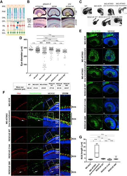
Figure 1. Knockdown of ataxin-3 in Zebrafish Embryos Alters Eye Size and Retinal Layer Organization.

(A) Schematic diagram of the mature vertebrate retina organization, which is similar in humans, mice, and zebrafish. RPE, retinal pigment epithelium; PhRs, photoreceptors; ONL, outer nuclear layer; OPL, outer plexiform layer; INL, inner nuclear layer; IPL, inner plexiform layer; GCL, ganglion cell layer; R, rod; C, cone.
(B) atxn3 is highly expressed in different retinal layers in zebrafish larvae (mRNA in situ hybridization on retinal cryosections at 7 days post-fertilization [dpf]). Sense atxn3 (negative) and antisense crx (positive) riboprobes were used as controls. Magnification, 20×; zoom, 40×.
(C) In vivo imaging of embryos (72 hpf) shows that microinjection of MO-ATXN3 (morpholino against atxn3) compared to MO-SCR (scrambled, negative control) causes alterations in the eye (white double-head arrows indicate the diameter) and head size depending on the level of atxn3 knockdown (low or high). The decrease in eye size was rescued when MO-ATXN3 was co-injected with human ATXN3 WT Q22 mRNA, but not as much with ATXN3 C14A (catalytically inactive mutant) or ATXN3 Q80 (MJD mutant). Scale bar, 50 μm.
(D) Quantification and comparison of eye size in the different embryo groups. A black line indicates the mean eye size in each group (n = 100 independent embryos per group). Mann-Whitney test (**p < 0.01, ***p < 0.001; n.s., non-significant).
(E) Extensive microtubule disorganization and defective formation of the retinal structures are observed in the retinas of high knockdown MO-ATXN3 morphants compared to MO-SCR (control) embryos, as shown by acetylated α-tubulin (green). Phenotypic rescue in retinal structures was observed after co-injecting morpholinos with human ATXN3-derived mRNAs, as indicated. Scale bar, 20 μm.
(F) Knockdown MO-ATXN3 zebrafish embryo retinas show elongation of the PhR outer segment (OS) and mislocalization of opsins compared to MO-SCR retinas (see zoom for detail, black bars on the right side indicate the OS length). This altered rod phenotype in MO-ATXN3 morphants is successfully rescued with co-injection of ATXN3 WT and C14A mRNAs. Rods are detected with rhodopsin (green) and cones with peanut agglutinin (PNA; red). White arrows indicate ectopic expression of opsins in cones and rods. Scale bar, 10 μm. Nuclei were counterstained with 4′,6-diamidino-2-phenylindole (DAPI) (blue).
(G) Rod OS (ROS) length measurements in morphants and rescued embryos (Mann-Whitney test; *p < 0.05; **p < 0.01; n.s., non-significant) (n = 3–4 images per embryo, 5 embryos per group).
